Decoding the Science of Glioma
- eftychiath
- Apr 19, 2022
- 7 min read
Updated: Oct 31, 2025
Abstract
Glioma is a malign brain tumor that begins in the glial cells and affects brain tissue (uncontrolled glial cells division). It is split in stages depending on its malignancy and affects 7 out of 100,000 patients yearly. In glioma, which is divided into gliosarcoma and glioblastoma, glial cells gain the characteristics of astrocytes and as time proceeds, grow uncontrollably, forming tumors. The main causes are old age, ionizing radiation and family history, while the symptoms include severe headaches, nausea and changes in one’s cognitive ability and behavior. Moreover, Glioma is diagnosed through an MRI, CT scan or Biopsy and treatment includes chemo and radiational therapy as well as surgical removal.
Introduction-Origins
Brain cancer could appear in specific types and takes place in the glial cell. One of the most common cancers in the form of an intracranial tumor, especially in adults is the glioma, which is split in the gliosarcoma and glioblastoma. In a global context, roughly 7 out of 100,000 patients yearly get the corresponding diagnosis (Tamimi & Juweid, 2017). Some of the first documented reports of gliomas were filed in Britain in 1800 and 1804. It is worth mentioning that during 1962 the foundations for the updated diagnosis and classification of gliomas were set (Stoyanov & Dzhenkov, 2018). Despite the passing of many years, a cure hasn’t been found, while the time of survival for diagnosed individuals continues to be 14-16 months (Aaron Cohen-Gadol, 2023).
Mechanism
The glioma identifies as a ‘’malignant’’ tumor. It is worth mentioning that gliomas grow quickly and are ranked from I to IV in accordance with the World Health Organization malignancy scale (benign tumor to malignant) (Marquet, Dameron, Saikali, Mosser & Burgun, 2007). The glioma relates to the procedure in which an unspecified cell, that looks like a glial cell gains the attributes of an astrocyte, a peculiar nerve cell which balances the environment in which it is housed, while at the same time, contributes to the function and development of neurons. Later, the glioma evolves into a group of masses, a tumor. Consecutively, after the tumor expands, it pressures the brain tissue, resulting to symptoms of the disease (Cancer Research UK, 2023). For instance, a frontal lobe tumor would include behavioral alternations, while a temporal lobe would come with discernable speech issues (Tamimi & Juweid, 2017).
The Two Types
The two main divisions of gliomas are the glioblastoma and gliosarcoma. The glioblastoma is a brain tumor which develops rapidly and is found to be very aggressive. The specific form of cancer starts in the astrocytes and afflicts the neighboring brain tissue and usually doesn’t reach organs which are further away. (Mayo Clinic Staff, 2023). Glioblastomas are evident most of the time in the cerebral hemispheres, particularly, in the brain’s frontal and temporal lobes (American Association of Neurological Surgeons, 2023). Furthermore, the gliosarcoma is also a tumor that occurs in the nervous system. It is one of the most common form of gliomas (National Cancer Institute, 2021). The main difference between glioblastoma and gliosarcoma is that gliosarcoma’s expansion pattern consists of both glial cells that support neurons as well as sarcomatous parts which have no connection with glioblastomas. In addition, gliosarcomas negatively affect connective tissues a main characteristic of sarcomas which isn’t evident in the case of glioblastomas (Fransen, Broholm, Larsen, Grunnet, Moller, Skovard & Michaelsen, 2019)(Rose, 2022).
Causes
There are several factors which should be taken into consideration while inspecting the causes of a glioma. To begin with, one of the most frequent reasons for the diagnosis of glioma is old age. Specifically, gliomas are more frequent in adults of 45-65 years. However, it has proven to affect individuals of any age. Another factor that should be conversed is exposure to radiation. To be precise, individuals who have faced a specific form of radiation, named ‘’ionizing’’ radiation have more chances of acquiring a brain tumor. Moreover, family history plays a significant role in determining the reasons for the appearance of the disease. It is yet under investigation, weather children, whose family members have suffered from glioma are more prone to getting it themselves (American Society of Clinical Oncology, 2023). Also, slight changes in the DNA of glial cells could trigger a malign brain tumor, while obesity increases one’s chances of establishing a tumor as such (Aaron Cohen-Gadol, 2023). Other detrimental factors include the sex of the person, as women are less likely than men to be diagnosed with glioma. Lastly, ethnicity is an agent that is related to the emergence of a glioma. For example, people who live in northern Europe are more susceptible to the illness than habitants of Japan (American Society of Clinical Oncology, 2023).
Signs and Symptoms
It is fact that every illness defines its presence through some symptoms. Firstly, one of the first symptoms portrayed by patients is a strong headache which is more intense in especially in the morning. Typically, glioma headaches become more recurrent and irresponsive to pain relieving medicine. Patients have referred to persistent vomiting and nausea. In addition, memory loss is observed. It should be stated that doctors have recognized mental confusion, deteriorating brain function such as problems with processing and comprehending information, as well as behavioral/personality changes and increased irritability in brain cancer patients. In some cases, there are visual difficulties, such as double or blurred vision, speech problems and a hard time with movement. Another common symptom is seizures, which is an abrupt and unexpected
outbreak of electrical activity inside the brain, which makes the patient tremor and move spasmodically/convulsively (National Health Service, 2023).
Diagnosis
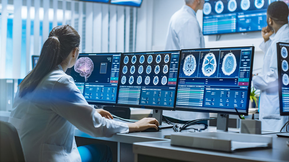
There are different procedures that help diagnose cancerous tumors such as the glioma. Most of the time, Magnetic resonance imaging is suggested (National Institute of Biomedical Imaging and Bioengineering, n.d.). In an MRI, glioma appears as a mass which is of great size and has necrosis as well as distinct edema (Toh & Castillo, 2017). The tumor also exerts intracranial pressure towards the brain tissues and is characterized by heterogenous enhancement (non-uniform) (National Library of Medicine, 2022). Additionally, there is also the computed tomography which provides doctors with 3-dimensional imaging. In a CT scan a tumor would appear to be white, making it stand out and more easily detectible (Markman, 2022). In rare situations, a biopsy is proposed, which is the removal tissue of the brain which is later put under a microscope and studied carefully (Mayo Clinic Staff, 2021).
Treatment
Most of the time, the procedure chosen, when looking for the urgent elimination a malignant tumor is surgical removal through a keyhole route, which is dependent on the tumor’s size and location. However, in early stages, one could follow chemotherapy and radiation therapy to kill cancer cells (Mayo Clinic Staff, 2023)(Cancer Research UK, 2023).
Conclusion
Taking all the above into consideration, it is apparent that the specific disease requires more study and medical research, as currently there is no effective, while people of all ages should commit to taking regular checkups, in order for it to be spotted in an early stage and be treated more effectively.
References
Aaron Cohen-Gadol M.D. “Long-Term Survivors of Glioblastoma: Expert Surgeon.” Aaron Cohen-Gadol MD, MSc, MBA, 7 Jan. 2023, www.aaroncohen-gadol.com/patients/glioma/survival/long-term-survivors#:~:text=Although%20the%20average%20life%20expectancy,than%2020%20years%20after%20diagnosis. Accessed 6 July 2023.
American Society of Clinical Oncology. “Brain Tumor - Risk Factors.” Cancer.Net, 31 May 2023, www.cancer.net/cancer-types/brain-tumor/risk-factors. Accessed 6 July 2023.
“Biopsy for Brain Tumours.” Biopsy for Brain and Spinal Cord Tumours | Cancer Research UK, 21 Apr. 2023, www.cancerresearchuk.org/about-cancer/brain-tumours/treatment/surgery/biopsy#:~:text=A%20biopsy%20means%20taking%20a,and%20won%27t%20feel%20anything. Accessed 6 July 2023.
Cleveland Clinic medical professional. “Glioma: What Is It, Causes, Symptoms, Treatment & Outlook.” Cleveland Clinic, 2023, my.clevelandclinic.org/health/diseases/21969-glioma#:~:text=Age%3A%20Gliomas%20are%20most%20common,common%20in%20men%20than%20women. Accesed 6 July 2023.
“CT Scan for Cancer Detection & Treatment.” Edited by Maurie Markman, City of Hope, 2 Mar. 2022, www.cancercenter.com/diagnosing-cancer/diagnostic-imaging/ct-scans#:~:text=Cancer%20cells%20take%20up%20the,important%20when%20making%20a%20diagnosis. Accessed 6 July 2023.
Frandsen, Simone, et al. “Clinical Characteristics of Gliosarcoma and Outcomes from Standardized Treatment Relative to Conventional Glioblastoma.” Frontiers, 29 Nov. 2019, www.frontiersin.org/journals/oncology/articles/10.3389/fonc.2019.01425/full#:~:text=Clinical%20Characteristics%20of%20Gliosarcoma%20and%20Outcomes%20From%20Standardized%20Treatment%20Relative%20to%20Conventional%20Glioblastoma,-Simone%20Frandsen1&text=Background%3A%20Gliosarcoma%20(GS)%20is,both%20glial%20and%20sarcomatous%20components. Accessed 6 July 2023.
“Glioma.” Brain Tumours (Primary) | Cancer Research UK, 7 June 2023, www.cancerresearchuk.org/about-cancer/brain-tumours/types/glioma-adults#:~:text=Gliomas%20are%20cancerous%20brain%20tumours,gliomas%20grow%20faster%20than%20others. Accessed 6 July 2023.
“Gliosarcoma Diagnosis and Treatment.” National Cancer Institute, 30 Sept. 2021, www.cancer.gov/rare-brain-spine-tumor/tumors/gliosarcoma#:~:text=Gliosarcoma%20is%20a%20primary%20central,removed%20during%20surgery%2C%20if%20possible. Accessed 6 July 2023.
“Glioma & Glioblastoma Symptoms, Diagnosis and Treatment.” Pacific Brain Tumor Center, 5 May 2023, www.pacificneuroscienceinstitute.org/brain-tumor/conditions/gliomas/#:~:text=The%20initial%20optimal%20treatment%20for,upon%20tumor%20location%20and%20size. Accessed 6 July 2023.
“Gliomas.” JHM, 11 Feb. 2022, www.hopkinsmedicine.org/health/conditions-and-diseases/gliomas#:~:text=Scans%20of%20the%20brain%3A%20Magnetic,for%20examination%20under%20a%20microscope. Accessed 6 July 2023.
“Glioblastoma Multiforme.” Edited by Jigisha P Thakkar et al., AANS, 2023, www.aans.org/en/Patients/Neurosurgical-Conditions-and-Treatments/Glioblastoma-Multiforme#:~:text=It%20invades%20the%20nearby%20brain,temporal%20lobes%20of%20the%20brain. Accessed 6 July 2023.
“Glioma - Symptoms, Causes, Treatment: Nord.” National Organization for Rare Disorders, 13 Aug. 2019, rarediseases.org/rare-diseases/glioma/. Accessed 6 July 2023.
MD Anderson Cancer Center, and Lauren Rose. “Glioma vs. Glioblastoma: What’s the Difference?” MD Anderson Cancer Center, 18 Feb. 2022, www.mdanderson.org/cancerwise/glioma-vs--glioblastoma--what-is-the-difference-in-these-brain-tumors-treatment-diagnosis.h00-159537378.html#:~:text=A%20glioma%20is%20one%20of,nerve%20endings%20in%20the%20brain.Accessed 6 July 2023.
Markman, Maurie. “CT Scan (CAT Scan) for Cancer Detection & Treatment.” City of Hope, 2 Mar. 2022, www.cancercenter.com/diagnosing-cancer/diagnostic-imaging/ct-scans#:~:text=Cancer%20cells%20take%20up%20the,cancerous%20lesion%2C%20including%20nearby%20organs. Accessed 6 July 2023.
Mayo Clinic Staff. “Glioma.” Mayo Clinic, 10 Jan. 2023, www.mayoclinic.org/diseases-conditions/glioma/symptoms-causes/syc-20350251. Accessed 6 July 2023.
Mefsin, Fassil B, and Mohammed A Al-Dhahir. “Gliomas - StatPearls - NCBI Bookshelf - National Center For for Biotechnology Information”, Gliomas, 2023, www.ncbi.nlm.nih.gov/books/NBK441874/. Accessed 6 July 2023.
National Health Service. “Chemotherapy.” NHS Choices, 25 May 2023, www.nhs.uk/conditions/chemotherapy/#:~:text=Chemotherapy%20is%20a%20cancer%20treatment,and%20spreading%20in%20the%20body. Accessed 6July 2023.
National Institute of Biomedical Imaging and Bioengineering. “Computed Tomography (CT).” National Institute of Biomedical Imaging and Bioengineering, June 2022, www.nibib.nih.gov/science-education/science-topics/computed-tomography-ct#:~:text=The%20term%20“computed%20tomography%2C”,images%2C%20or%20“slices.” Accessed 6 July 2023.
National Institute of Biomedical Imaging and Bioengineering. “Magnetic Resonance Imaging (MRI).” National Institute of Biomedical Imaging and Bioengineering, www.nibib.nih.gov/science-education/science-topics/magnetic-resonance-imaging-mri#:~:text=MRIs%20employ%20powerful%20magnets%20which,pull%20of%20the%20magnetic%20field. Accessed 6 July 2023.
Prabhu, Vikram C. “Glioblastoma Multiforme.” Edited by Jigisha P Thakkar and Pier Paolo Peruzzi, American Association of Neurological Surgeons, www.aans.org/en/Patients/Neurosurgical-Conditions-and-Treatments/Glioblastoma-
Tamimi, Ahmad Faled, and Malik Juweid. “Epidemiology and Outcome of Glioblastoma - Glioblastoma - NCBI Bookshelf.” National Library of Medicine, 17 Sept. 2017, www.ncbi.nlm.nih.gov/books/NBK470003/. Accessed 6 July 2023.
Toh, C H, and M Castillo. “Early-Stage Glioblastomas: Mr Imaging-Based Classification and Imaging Evidence of Progressive Growth.” AJNR. American Journal of Neuroradiology, Feb. 2017, www.ncbi.nlm.nih.gov/pmc/articles/PMC7963843/#:~:text=It%20typically%20appears%20as%20a,heterogeneous%20contrast%20enhancement%20when%20diagnosed. Accessed 6 July 2023.


Comments